
Triplet V1
首个面向病理多尺度推理的基础大模型
Triplet V1 以跨倍率推理为核心能力,让模型像病理医生一样完成视觉规划、逐级验证与结论收敛,并输出可追溯的证据链。
Benchmark 结果
78.1%
跨尺度 VQA 领先表现
在 4,810 道 Triplet-VQA 测试题上,较对比模型中的最佳结果高 18.2 个百分点。
多中心临床验证
10.9–25.9%
报告效率提升
四中心配对研究显示报告时间节省 10.9%–25.9%;九中心临床诊断报告辅助中,编辑修订字段修改率为 29.3%。
设计与功能优势
10× → 40× → 200×
可解释、可交互的推理
逐级连接组织结构与细胞学证据,完整呈现推理路径,并支持医生审核、修正与回溯。
为什么是多尺度推理
病理诊断不是看一张图,而是先低倍扫全局,再中倍定位结构,最后高倍确认细胞学。Triplet 把这条跨倍率流程定义为模型的核心能力目标,并据此设计训练与评测。
全景扫描:识别组织整体结构与异常区域分布,为多尺度推理提供起点。
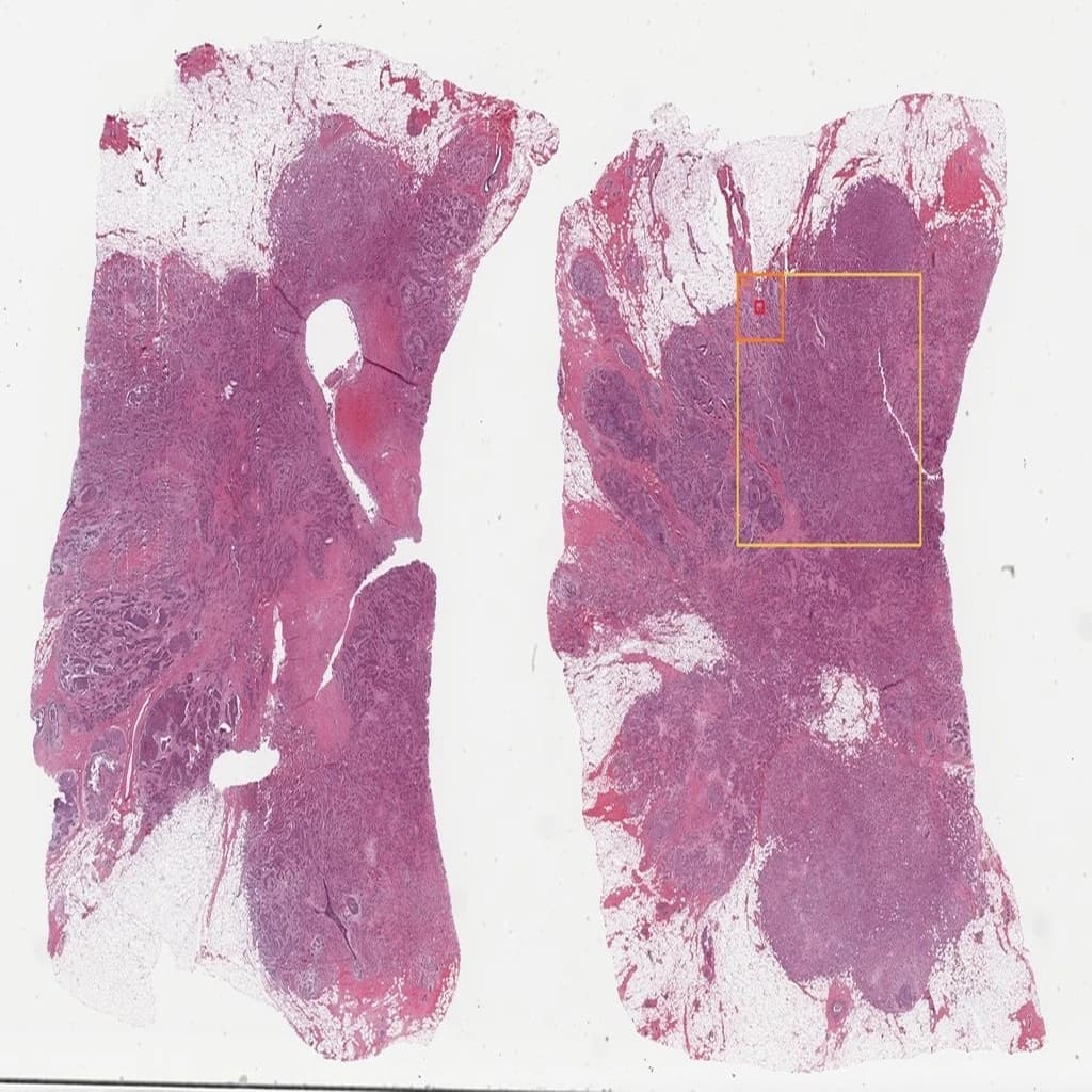
低倍镜下可见导管/腺泡结构紊乱伴间质纤维化及细胞密集,提示组织结构破坏;中倍镜显示上皮细胞排列紊乱、核增大深染、基底膜不完整及肌上皮缺失,提示上皮异型性进展;高倍镜下核异型性显著、核浆比增高、染色质不均及病理性核分裂象,符合恶性肿瘤细胞特征。
导管/腺泡结构紊乱,间质纤维化,细胞密集
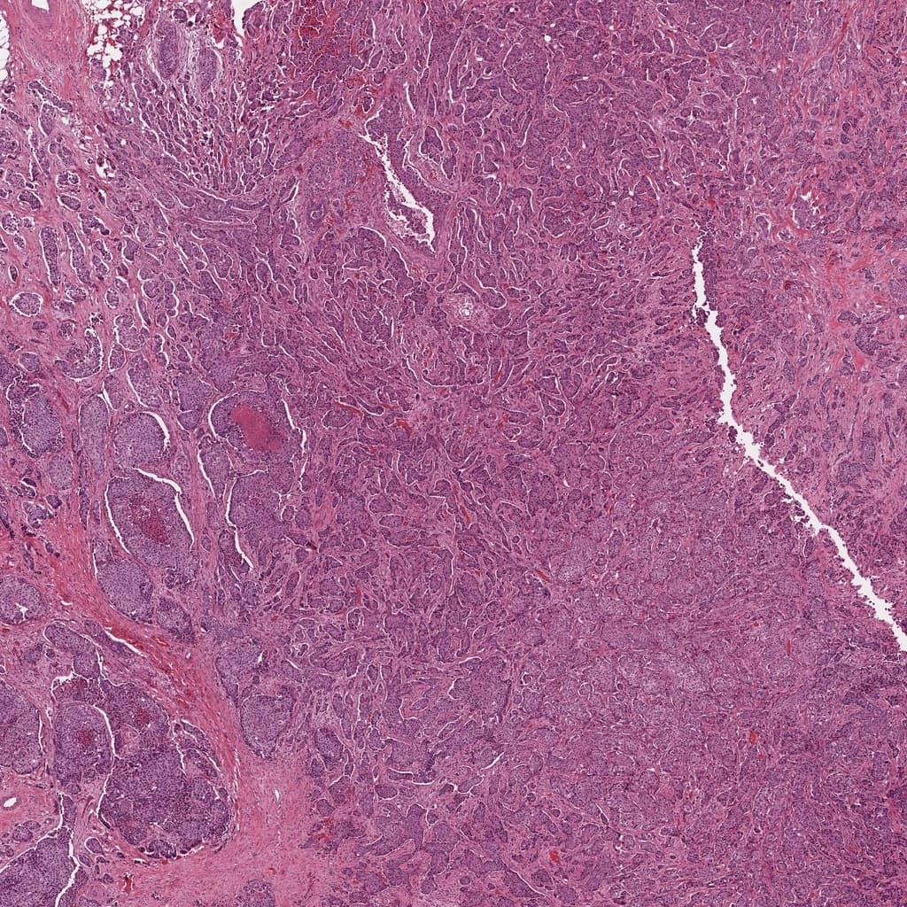
在10×放大下,导管/腺泡排列不规则,部分区域呈背靠背结构,间质比例显著增加并伴硬化性改变,细胞核密集、大小不一,局部可见轻度炎性浸润,整体结构拥挤。
上皮细胞排列紊乱,核增大深染,核仁明显,胞浆减少,基底膜不完整伴肌上皮缺失
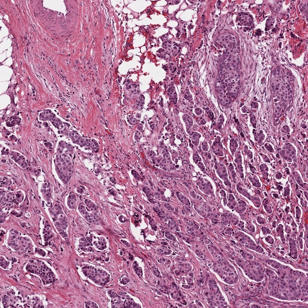
在20×放大下,组织结构不规则,上皮细胞呈巢状或条索状浸润性生长,细胞极性丧失,核体积增大、深染、大小不一,核仁明显,胞浆稀少,间质纤维化伴炎细胞浸润,基底膜连续性中断,肌上皮层缺失。
细胞核异型性显著,核浆比增高,染色质深染且分布不均,核仁明显,可见病理性核分裂象
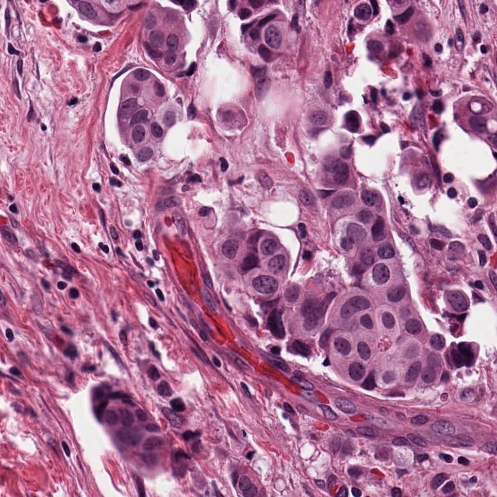
在40×放大下,组织结构紊乱,细胞排列密集且极向丧失,核体积增大、形态不规则、染色质深染且分布不均,核仁显著,核浆比增高,间质纤维化伴炎细胞浸润,可见多处病理性核分裂象。
Triplet 大模型的技术优势
跨倍率推理,全流程可解释
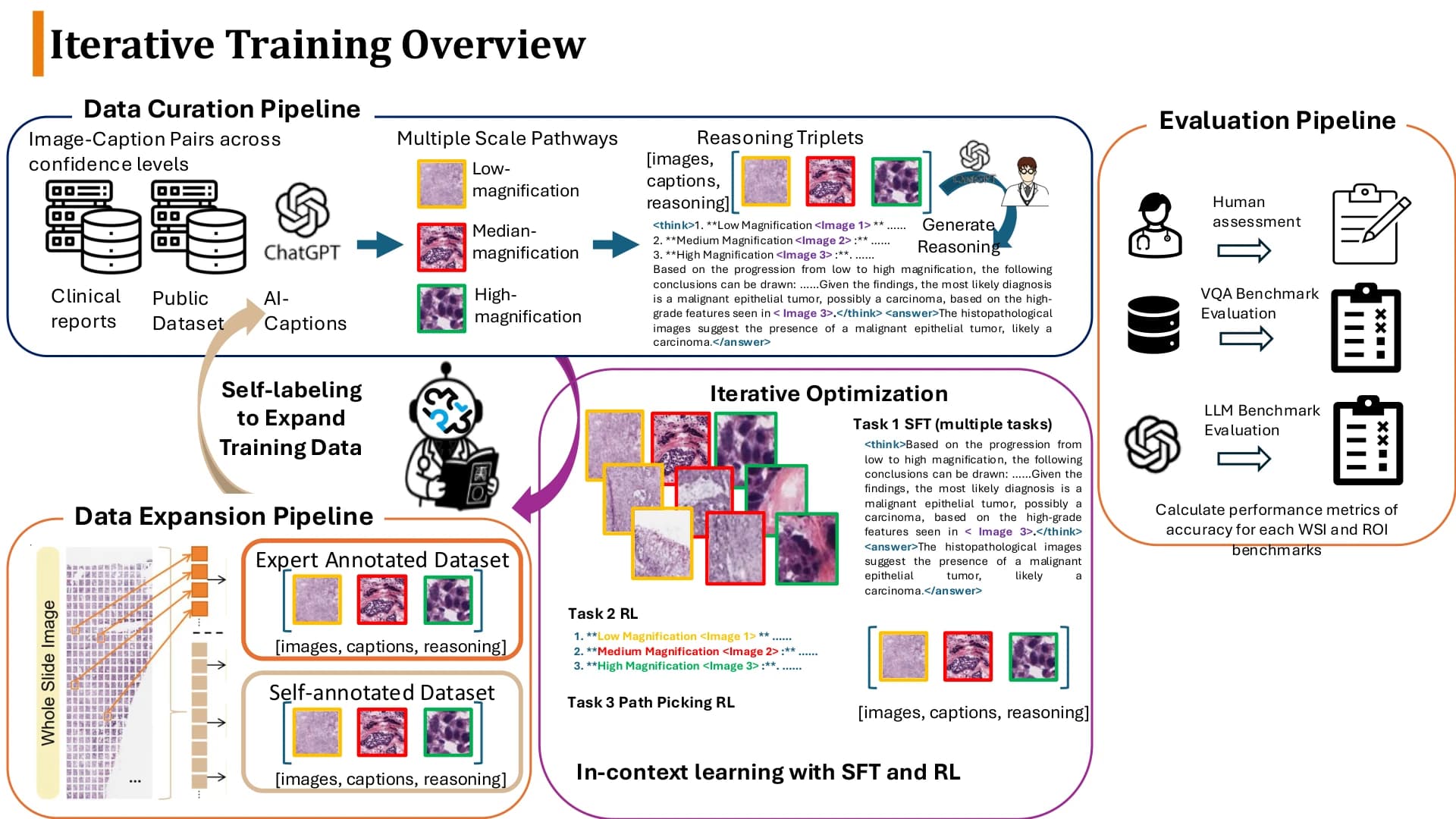
Triplet 特有多尺度可解释证据链训练,逐步循证推理,构建临床可审核的推理表达。
推理可交互,证据可回溯
逐级呈现从低倍组织结构到高倍细胞学证据,医生可随时修正与回溯,形成闭环推理。
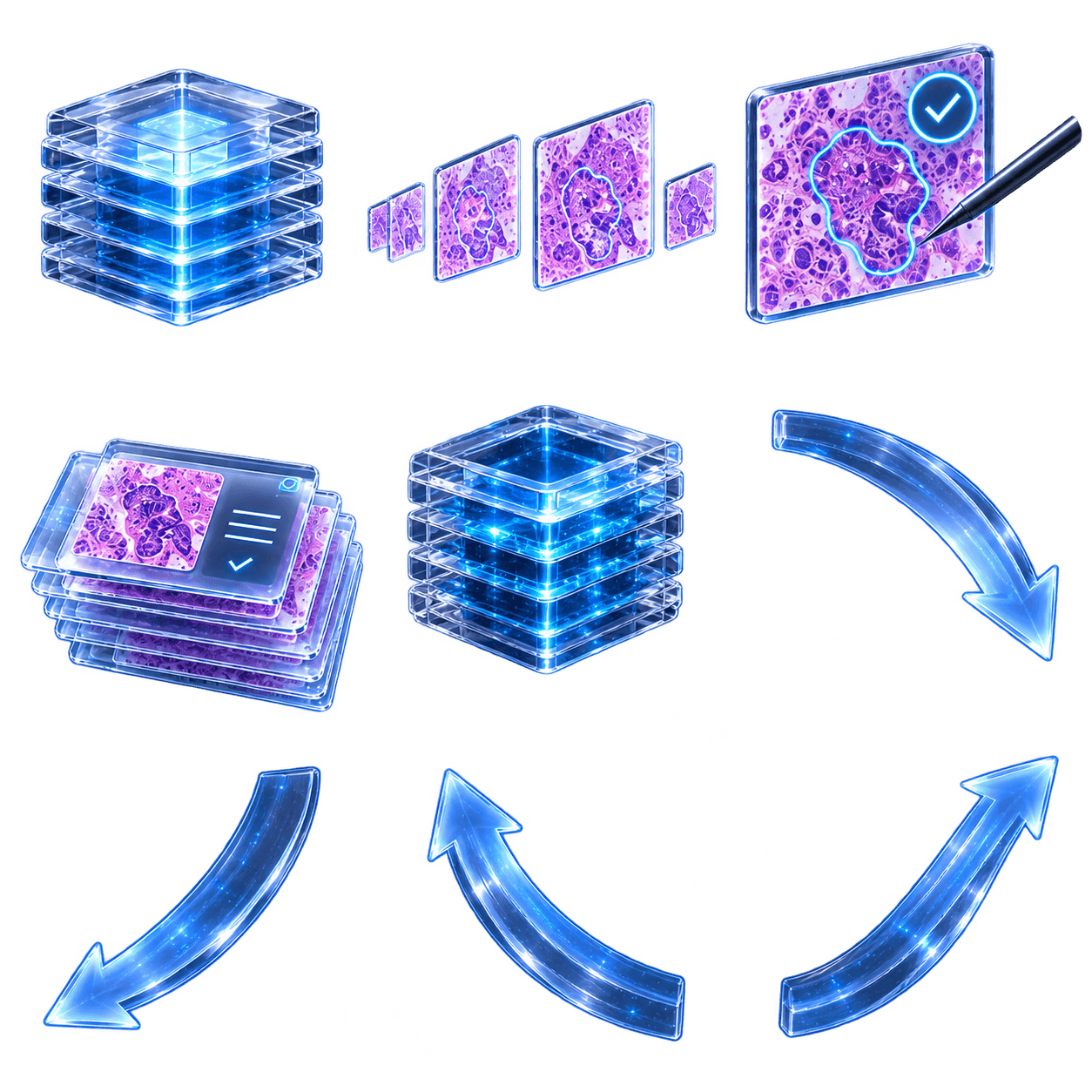
人在回路数据飞轮
人机循环数据标注,AI 生成、医生审核并返哺训练,构建数据与模型能力持续增长的闭环。
多区域异质性理解
学习一条 Path 上多个 ROI 的前后文关系,理解该路线对整张 WSI 的诊断意义。
结构化输出(适合临床复核)
每个倍率输出放大动机 Reason、图像描述 Caption,并形成跨尺度推理链与总结。
可选中英输出
支持中英表达,但主叙事聚焦在跨倍率推理与证据链能力。
核心机制:Triplet 三元结构
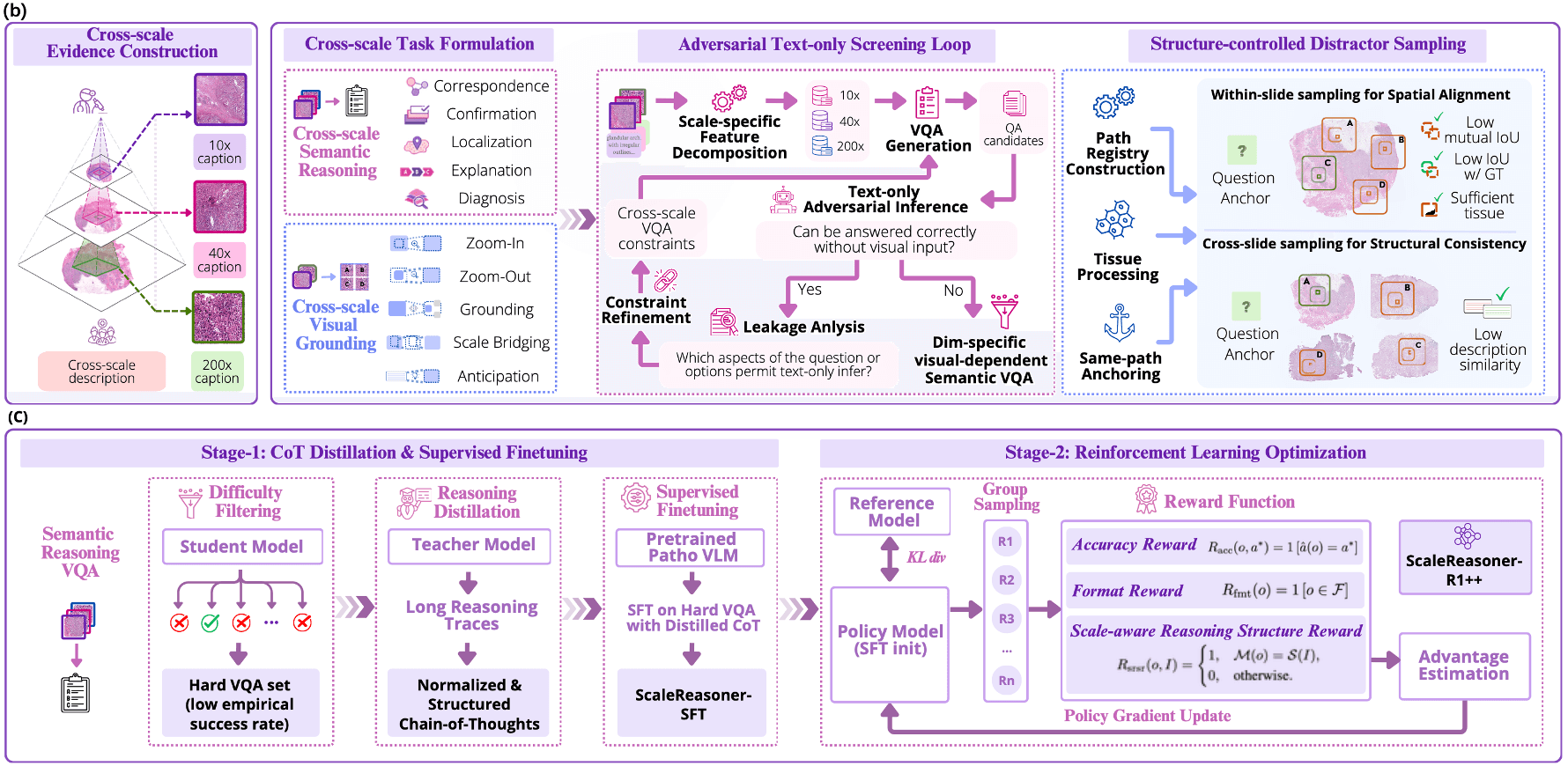
Triplet 用三元组数据结构组织学习:多尺度图像 + 选择理由与病理特征 + 路线的诊断意义与发现。每条路线是一条完整的跨尺度 Path(10x → 40x → 200x)。
从数据构建到部署使用
从高质量、多尺度、可解释的数据构建,到在 PuzzleVision 中完成区域分析与全切片推理,Triplet 覆盖模型开发与实际应用的完整流程。
将 Triplet 接入 PuzzleVision 阅片工作台,在真实 WSI 流程中完成区域分析、跨倍率证据整合与全切片推理。
训练与评测
SFT 让模型理解证据路线;RL 让"会推理"可以被可靠衡量。
SFT:让模型理解证据路线
基于多尺度每个区域构建跨尺度 Path(10x → 40x → 200x),逐层学习 Reason / Caption / 推理链 / 总结,让模型先学会"为什么放大、看到了什么、如何归纳"。
RL:让"会推理"被可靠衡量
采用 leakage-aware 机制抑制文本捷径:先做 text-only 对抗推断筛查,若不看图也能答对则判定泄漏并迭代修正。随后用 GRPO 对齐,奖励答案正确性与跨尺度推理一致性,并约束输出格式稳定。
Benchmark
模型能力评测
以海量公开数据与多中心私有数据为 benchmark,在多图推理与单图分析任务上与 SOTA 模型进行全面对比。
Triplet-VQA 总体准确率
78.1%
较对比模型中表现最佳的 Lingshu-7B(59.9%)高 18.2 个百分点
评测规模
4,810 题 / 15 个器官系统
精确匹配(exact-match)准确率;五个问题类型各 962 题
Overall accuracy
Triplet-VQA 总体排名
五类问题精确匹配准确率的平均值,比较 7 个代表性模型。
五项能力对比
| Model | 对应性 | 确认性 | 定位 | 解释 | 诊断 | Mean |
|---|---|---|---|---|---|---|
| TRIPLET | 70.2 | 84.4 | 88.4 | 63.4 | 84.2 | 78.1 |
| Lingshu-7B | 53.5 | 67.4 | 73.7 | 57.6 | 47.6 | 59.9 |
| CLOVER | 48 | 64.5 | 79.2 | 48.7 | 51.7 | 58.4 |
| QoQ-Med-VL-7B | 49.7 | 54.7 | 68.9 | 41.5 | 57.1 | 54.4 |
| Patho-R1 | 32.5 | 35.9 | 63 | 44.9 | 52.6 | 45.8 |
| PRISM2 | 32.1 | 46.3 | 33 | 31.4 | 25.1 | 33.6 |
| Quilt-LLaVA | 24.6 | 18.7 | 41.4 | 28.4 | 26.6 | 27.9 |
查看 VQA 案例与各模型推理思路+
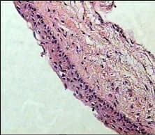
题目
Based on the cellular morphology, which layer of stratified squamous epithelium is most prevalent on the left side of the image? Translation 基于细胞形态,哪一层的复层鳞状上皮在图像的左侧最常见?
模型回答对比
案例数据来源于 Triplet-V1 论文,展示不同模型在跨尺度推理任务上的回答对比。
Clinical studies
临床实验部署
从报告速度、报告质量和报告修订负担三个互补终点评估人机协作价值。
01
更快完成报告
节省 10.9 – 25.9%
同一医生、同一 WSI 的配对计时,四个中心的节省区间
02
改善报告质量
3.97 → 4.70
低年资医生组报告总体评分;75% 的配对盲评更偏好人机协作
03
降低报告修订负担
29.3%
编辑修订字段修改率,比 Qwen3.5-plus 少 13.6 个百分点
01 · Speed
更快完成报告
同一医生、同一 WSI,手工与人机协作配对计时。
节省时间范围

福建省肿瘤医院
FJCH · n=63
P<0.1 · 57.1% 病例更快

山西省肿瘤医院
SXCH · n=60
P<0.11 · 56.7% 病例更快

宁波病理中心
NBPC · n=76
P<0.001 · 75% 病例更快

上海六院
SSPH · n=49
P=0.001 · 73.5% 病例更快
共 4 个中心、248 例配对 WSI;双侧配对 Wilcoxon 检验。宁波病理中心与上海六院达统计学显著。福建省肿瘤医院展示值已按部署延迟敏感性分析调整。
02 · Quality
改善报告质量
两个独立中心、两组不同资历的医生;每组报告由一位独立资深评审者盲评。
山西省肿瘤医院
低年资医生组 · n=60
1–5
诊断准确性
P=0.081
镜下描述完整性
P=7.1e-11
描述—诊断一致性
P=6.6e-07
语言与术语
P=1.8e-09
总体印象
P=5.3e-09
配对盲评偏好
福建省肿瘤医院
资深医生组 · n=59
1–5
诊断准确性
P=0.317
镜下描述完整性
P=0.034
描述—诊断一致性
P=0.549
语言与术语
P=0.034
总体印象
P=1
配对盲评偏好
低年资组总体评分 3.97 → 4.70,75% 偏好人机协作。资深组总体评分 4.29 → 4.29(P=1);语言与术语评分由 4.73 降至 4.53。未达到显著不等同于等效或非劣。
03 · Revision burden
降低报告修订负担
同一 WSI 的三份模型生成报告以盲法随机顺序审核;编辑修订字段修改率越低,表示医生复核负担越小。
9 中心 · 457 张 WSI · 6 位病理医生
复核时间(441 条完整记录)
vs Qwen3.5-plus
26.3s 更快
95% CI -37.9 to -21.3s · Holm P=5.1e-14
vs Gemini 3
9.5s 更快
95% CI -18.2 to -3.1s · Holm P=0.002
平均复核时间:TRIPLET 138.3 秒,Gemini 3 154.4 秒,Qwen3.5-plus 170.5 秒。
查看 9 个中心的修改率差异与置信区间+
| Center | n | Baseline | Difference (pp) | 95% CI | Holm P |
|---|---|---|---|---|---|
| 辽宁省肿瘤医院 | 49 | Qwen3.5-plus | -6.8 | -13.5 to -0.4 | 0.199 |
| 辽宁省肿瘤医院 | 49 | Gemini 3 | -3.2 | -8.9 to 2.5 | 0.584 |
| 上海六院 | 52 | Qwen3.5-plus | -13.5 | -19.2 to -7.6 | 0.001 |
| 上海六院 | 52 | Gemini 3 | -2.9 | -8.3 to 2.8 | 0.097 |
| 上海市一医 | 50 | Qwen3.5-plus | -4.7 | -12.9 to 3.2 | 0.712 |
| 上海市一医 | 50 | Gemini 3 | +2.9 | -4 to 9.5 | 0.712 |
| 山西省肿瘤医院 | 68 | Qwen3.5-plus | -7 | -10.9 to -3.1 | 0.002 |
| 山西省肿瘤医院 | 68 | Gemini 3 | +0.9 | -2.9 to 4.7 | 0.664 |
| 福建省肿瘤医院 | 71 | Qwen3.5-plus | -21.7 | -26.6 to -16.7 | 2.4e-09 |
| 福建省肿瘤医院 | 71 | Gemini 3 | -12.9 | -17.7 to -8.3 | 1.0e-05 |
| 北京协和医院 | 43 | Qwen3.5-plus | -27.7 | -34.3 to -21.2 | 5.4e-07 |
| 北京协和医院 | 43 | Gemini 3 | -16 | -21.8 to -10.3 | 2.2e-05 |
| 宁波病理中心 | 78 | Qwen3.5-plus | -18.9 | -23 to -14.9 | 1.4e-10 |
| 宁波病理中心 | 78 | Gemini 3 | -12.4 | -16.3 to -8.5 | 3.9e-07 |
| 北京胸科医院 | 26 | Qwen3.5-plus | -12.7 | -16.8 to -8 | 7.9e-04 |
| 北京胸科医院 | 26 | Gemini 3 | -9.8 | -15.1 to -4.2 | 0.007 |
| 上海东方肝胆医院 | 20 | Qwen3.5-plus | +3.9 | -5.8 to 13.1 | 0.681 |
| 上海东方肝胆医院 | 20 | Gemini 3 | +5.4 | -3 to 14.2 | 0.681 |
| 全部中心 | 457 | Qwen3.5-plus | -13.6 | -15.6 to -11.5 | 3.1e-28 |
| 全部中心 | 457 | Gemini 3 | -6.2 | -8.1 to -4.3 | 4.9e-10 |
TRIPLET 相比 Qwen3.5-plus 在 6 个中心显著更低,相比 Gemini 3 在 4 个中心显著更低;EHBH 方向相反但不显著。
Extended analysis
更多实验与分析
进一步评估 ROI 与 WSI 分类、自主诊断、置信度复核及五轮数据标注质量。
更多 Benchmark:ROI / WSI 分类、消融与 LLM 评分+
实验发现
跨尺度、同病灶对齐的 Triplet 监督带来的收益并非仅来自“看见更多倍率”:独立图文对训练的平均准确率为 58.6%,而完整 TRIPLET 达到 78.1%。这一结构同时迁移到单图问答及 ROI / WSI 分类,并在自由文本评估中取得最高的感知、推理与结论评分。
ROI 分类(10 个数据集)
| Model / group | CCRCC | Chaoyang | BreakHis | Herlev | GasHisSDB | LC25000 | PAIP | HMU-GC-HE | CRC-100K | WSSS4LUAD | Mean |
|---|---|---|---|---|---|---|---|---|---|---|---|
| Lingshu-7B · ACC | 55.6 | 53 | 56 | 17.9 | 54 | 48.3 | 52.6 | 35.3 | 52.9 | 42.3 | 46.8 |
| Lingshu-7B · macro-F1 | 37.3 | 25.6 | 47.4 | 8.9 | 52.6 | 40.8 | 49.7 | 29.8 | 43.3 | 37.7 | 37.3 |
| Quilt-LLaVA · ACC | 34 | 21 | 58.4 | 12.8 | 53.8 | 35 | 47.6 | 14.1 | 8 | 33.1 | 31.8 |
| Quilt-LLaVA · macro-F1 | 21.4 | 15.6 | 32.6 | 9.5 | 53.1 | 20.8 | 31.7 | 9 | 7 | 33.1 | 23.4 |
| CLOVER · ACC | 43.3 | 45.9 | 49.7 | 24.8 | 57.7 | 61.3 | 56 | 29.6 | 34.9 | 52.2 | 45.5 |
| CLOVER · macro-F1 | 30.3 | 32.7 | 19.4 | 17.7 | 37.6 | 46.9 | 54.9 | 28.8 | 32.4 | 52.9 | 35.4 |
| QoQ-Med-VL-7B · ACC | 51.2 | 48.5 | 47.7 | 20.8 | 52.4 | 21.7 | 59.5 | 18.8 | 25.1 | 56.4 | 40.2 |
| QoQ-Med-VL-7B · macro-F1 | 24.9 | 28.8 | 30.3 | 9 | 50.8 | 24.9 | 64.8 | 15.2 | 23.7 | 41.8 | 31.4 |
| Patho-R1 · ACC | 63.9 | 54.8 | 58.4 | 18.2 | 43.6 | 78.1 | 55.1 | 34.8 | 62.8 | 57.5 | 52.7 |
| Patho-R1 · macro-F1 | 57.8 | 27.5 | 37.8 | 14.4 | 24.7 | 58.5 | 54.9 | 29.4 | 50.3 | 58.1 | 41.3 |
| PRISM2 · ACC | 65.7 | 35.2 | 69.8 | 22.6 | 46.1 | 62.7 | 74.6 | 26.5 | 57.1 | 28.1 | 48.8 |
| PRISM2 · macro-F1 | 49.2 | 29.7 | 68.9 | 10.2 | 45.3 | 53 | 60.9 | 22.7 | 43.7 | 19.2 | 40.3 |
| TRIPLET · ACC | 58.7 | 61.1 | 53.7 | 26 | 61.6 | 67.3 | 58.7 | 36.1 | 70 | 59.5 | 55.3 |
| TRIPLET · macro-F1 | 53.7 | 43.3 | 50.3 | 23.9 | 61.3 | 63.8 | 57.2 | 32.9 | 64.8 | 56.4 | 50.8 |
在 ROI 分类评测中,TRIPLET 平均 ACC 为 55.3%,Qwen3.5-VL 为 58.3%;PRISM2 在 PAIP、BreakHis、CCRCC 上高于 TRIPLET。
WSI 分类(3 个数据集)
| Model / group | CAMELYON-16 | BRACS | CPTAC-LUNG | Mean |
|---|---|---|---|---|
| Lingshu-7B · ACC | 44.8 | 12.8 | 53 | 36.9 |
| Lingshu-7B · macro-F1 | 40.2 | 2.9 | 39.2 | 27.5 |
| Quilt-LLaVA · ACC | 49.3 | 20.9 | 50.8 | 40.3 |
| Quilt-LLaVA · macro-F1 | 48.4 | 11.8 | 49 | 36.4 |
| CLOVER · ACC | 50.7 | 16.3 | 44.5 | 37.2 |
| CLOVER · macro-F1 | 50.7 | 12 | 29.6 | 30.8 |
| QoQ-Med-VL-7B · ACC | 43 | 23.3 | 48.5 | 38.3 |
| QoQ-Med-VL-7B · macro-F1 | 35.1 | 17.7 | 32.6 | 28.5 |
| Patho-R1 · ACC | 53.7 | 12.8 | 51.9 | 39.5 |
| Patho-R1 · macro-F1 | 42.1 | 5.1 | 39.9 | 29 |
| PRISM2 · ACC | 42.2 | 9.3 | 57.8 | 36.4 |
| PRISM2 · macro-F1 | 32.6 | 4.5 | 56.9 | 31.3 |
| TRIPLET · ACC | 57.8 | 24.4 | 53.2 | 45.1 |
| TRIPLET · macro-F1 | 48.6 | 18.4 | 53 | 40 |
Caption-mode 消融
| Model / group | 对应性 | 确认性 | 定位 | 解释 | 诊断 | Mean |
|---|---|---|---|---|---|---|
| TRIPLET | 70.2 | 84.4 | 88.4 | 63.4 | 84.2 | 78.1 |
| Triplet only | 52.6 | 52.4 | 78 | 44.7 | 74.8 | 60.4 |
| 10×–40×–200× (none removed) | 50.6 | 53.8 | 73.9 | 45.5 | 69 | 58.6 |
| 10×–40× (200× removed) | 47.4 | 55.4 | 76.3 | 46 | 65 | 58 |
| 10×–200× (40× removed) | 47.4 | 56.7 | 76.8 | 42.1 | 68.3 | 58.2 |
| 40×–200× (10× removed) | 53.1 | 59.9 | 77.4 | 47.8 | 66.4 | 60.9 |
LLM-as-judge(0–5 分)
| Model / group | Perception mean | SD | Reasoning mean | SD | Conclusion mean | SD |
|---|---|---|---|---|---|---|
| Patho-R1 | 3.53 | 0.91 | 3.91 | 0.67 | 3.86 | 1.57 |
| Lingshu-7B | 3.61 | 0.55 | 3.78 | 0.41 | 4.4 | 0.94 |
| HuatuoGPT-V-7B | 3.42 | 0.56 | 3.91 | 0.33 | 4.28 | 1.14 |
| Qwen3.5-VL-9B | 3.34 | 0.98 | 3.97 | 1.17 | 3.72 | 1.7 |
| Qwen3.5-plus | 3.29 | 1.13 | 3.94 | 1.21 | 3.49 | 1.92 |
| TRIPLET | 3.71 | 0.69 | 4.28 | 0.68 | 4.48 | 1.09 |
进一步研究:自主诊断(160 张 WSI,9 中心)+
实验发现
当模型获得病灶对齐的跨尺度 ROI 与整合摘要时,基本癌种准确率达到 90.6%–98.8%;仅依赖 WSI 缩略图时降至 48.1%–48.8%。这说明证据选择与组织本身是诊断流程的一部分;由于两种输入条件并不等价,该结果更适合作为分流与预筛研究依据,而非自动签发结论。
证据条件与模型
| Model / group | Accuracy (%) | 95% CI | Score / 100 |
|---|---|---|---|
| TRIPLET-guided ROI evidence · TRIPLET | 98.8 | 95.6–99.7 | 84.3 |
| TRIPLET-guided ROI evidence · Gemini 3 | 91.9 | 86.6–95.2 | 76.4 |
| TRIPLET-guided ROI evidence · Qwen3.5-plus | 90.6 | 85.1–94.2 | 78.7 |
| WSI thumbnail only · Qwen3.5-plus | 48.8 | 41.1–56.4 | 46.4 |
| WSI thumbnail only · Gemini 3 | 48.1 | 40.5–55.8 | 46.6 |
分中心基本癌种准确率
| Model / group | 辽宁省肿瘤医院 | 上海六院 | 上海市一医 | 山西省肿瘤医院 | 福建省肿瘤医院 | 北京协和医院 | 宁波病理中心 | 北京胸科医院 | 上海东方肝胆医院 |
|---|---|---|---|---|---|---|---|---|---|
| ROI evidence · TRIPLET | 100 | 100 | 100 | 100 | 100 | 100 | 100 | 100 | 80 |
| ROI evidence · Gemini 3 | 100 | 90 | 95 | 95 | 90 | 95 | 90 | 90 | 70 |
| ROI evidence · Qwen3.5-plus | 95 | 100 | 85 | 95 | 90 | 95 | 90 | 70 | 80 |
| Thumbnail · Qwen3.5-plus | 55 | 80 | 55 | 70 | 40 | 55 | 35 | 0 | 0 |
| Thumbnail · Gemini 3 | 55 | 75 | 50 | 80 | 40 | 50 | 35 | 0 | 0 |
分器官准确率
| Model / group | n | TRIPLET + ROI | Gemini 3 + ROI | Qwen + ROI | Gemini thumbnail | Qwen thumbnail |
|---|---|---|---|---|---|---|
| Breast | 46 | 100 | 93.5 | 97.8 | 71.7 | 65.2 |
| Colorectal | 22 | 100 | 100 | 95.5 | 100 | 100 |
| Hepatobiliary | 16 | 87.5 | 75 | 68.8 | 0 | 18.8 |
| Lung | 44 | 100 | 88.6 | 81.8 | 0 | 4.5 |
| Gastric | 32 | 100 | 96.9 | 100 | 68.8 | 65.6 |
病例按 existing_local_best 策略选择,不代表随机前瞻性队列。150 例采用 strict 规则,EHBH 10 例采用参考比对宽松规则,因此汇总为混合口径。
进一步研究:置信度与医生复核分流+
实验发现
置信度能够识别更复杂、更需要复核的病例:以 0.75 为阈值,53 例进入复核组,其中包括 15 / 20 例 cHCC-CCA 和全部 10 例 iCCA。该结果支持用置信度安排复核优先级,而不是将高置信度作为自动签发标准。
100 张肝胆 WSI
| Model / group | n | Strict accuracy (%) | Lineage-aware (%) |
|---|---|---|---|
| All WSIs | 100 | 64 | 87 |
| High confidence (≥0.75) | 47 | 80.85 | 97.87 |
| Review arm (<0.75) | 53 | 49.06 | 77.36 |
按参考诊断
| Model / group | WSIs | Patients | Strict (%) | Lineage-aware (%) | Mean confidence | Routed to review (%) |
|---|---|---|---|---|---|---|
| HCC | 70 | 70 | 85.71 | 92.86 | 0.74 | 40 |
| cHCC-CCA | 20 | 20 | 5 | 95 | 0.66 | 75 |
| iCCA | 10 | 2 | 30 | 30 | 0.65 | 100 |
混淆矩阵
| Model / group | Pred. HCC | Pred. cHCC-CCA | Pred. iCCA | Pred. other | Total |
|---|---|---|---|---|---|
| HCC | 60 | 5 | 1 | 4 | 70 |
| cHCC-CCA | 13 | 1 | 5 | 1 | 20 |
| iCCA | 7 | 0 | 3 | 0 | 10 |
ECE 0.0799;Brier score 0.216。高置信度(≥0.75)结果仍需病理医生审核,置信度仅用于复核排序。10 张 iCCA 来自 2 位患者。
进一步研究:五轮数据标注质量+
实验发现
五轮迭代中,放大理由修改率由 23.1% 降至 10.2%,描述修改率由 27.8% 降至 12.9%,平均质量评分由 3.84 升至 4.21。结果表明人机循环使候选标注逐步接近专家确认目标,同时最终监督信号仍由病理医生把关。
轮次趋势(共 21,853 条路径)
| Model / group | Scored paths | Reason edits (%) | Caption edits (%) | Mean quality | 5-star share (%) |
|---|---|---|---|---|---|
| Round 1 | 4498 | 23.15 | 27.8 | 3.841 | 23.34 |
| Round 2 | 5853 | 20.17 | 27.68 | 3.774 | 21.22 |
| Round 3 | 4826 | 14.21 | 22.05 | 3.978 | 31.19 |
| Round 4 | 4582 | 13.97 | 16.39 | 4.009 | 31.14 |
| Round 5 | 2094 | 10.22 | 12.91 | 4.213 | 44.41 |
评分分布
| Model / group | 1 star (%) | 2 stars (%) | 3 stars (%) | 4 stars (%) | 5 stars (%) |
|---|---|---|---|---|---|
| Round 1 | 4.36 | 5.78 | 14.63 | 51.89 | 23.34 |
| Round 2 | 4.65 | 8.12 | 13.63 | 52.38 | 21.22 |
| Round 3 | 3.46 | 5.47 | 12.06 | 47.82 | 31.19 |
| Round 4 | 3.12 | 5.33 | 10.21 | 50.2 | 31.14 |
| Round 5 | 1.96 | 3.68 | 9.93 | 40.02 | 44.41 |
标注员校准
| Model / group | Mean quality | Character modification (%) |
|---|---|---|
| Annotator 1 | 3.34 | 25.31 |
| Annotator 2 | 4.45 | 11.99 |
| Annotator 3 | 4.12 | 24.5 |
| Annotator 4 | 4.64 | 26.98 |
| Annotator 5 | 3.7 | 12.53 |
| Annotator 6 | 2.67 | 18.52 |
| Annotator 7 | 3.82 | 17.63 |
| Annotator 8 | 4.63 | 8.82 |
字符修改率是标注流程的字符级指标,与临床诊断报告辅助中的编辑修订字段修改率不同。八位标注员对相同模型输出的评分用于标注一致性校准,不作为语料过滤标准。
在线试用
上传或选择一张示例图像,体验 Triplet 的跨倍率推理输出。
图像输入
支持上传或使用预设图像
分析结果
接入Triplet:敬请期待
面向系统接入、工作流协作与研究版强化推理的三种形态。
Triplet API
把跨倍率推理能力接入你的系统:VQA、结构化描述、证据链输出、质控接口。
Triplet Agents(协作版)
面向工作流:区域选择 -> 证据汇总 -> 报告草拟 -> 人类医生监督闭环。
Triplet Reasoning Edition(研究/高级版)
基于 leakage-aware 数据构建 + GRPO 强化学习,进一步提升跨尺度一致性与可控输出。
FAQ
Triplet V1 的核心差异是什么?
跨倍率推理是第一能力。训练、数据与评测都围绕"证据链闭环"设计,不做单尺度黑箱判断。
你们的数据如何保证可信?
采用循环式 human-in-the-loop:AI 先产出,医生审核修正并反哺训练,持续提升数据质量与可追溯性。
Triplet 的数据来自哪里?
私有数据均来自经过伦理审核的多中心科研合作,并按照合规要求获取和使用;公开数据覆盖全球 100 余个公开数据集。
Triplet 的数据由谁标注与审核?
来自三甲医院的 100 余位病理医师参与数据标注与审核,通过专业复核持续保障标注的准确性、一致性与临床相关性。